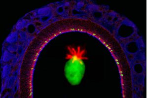
An image of main olfactory epithelium (MOE) from a ChAT-GFP mouse. Cholinergic TRPM5-expressing microvillous cells (TRPM5-MCs) express ChAT (GFP) and are located at superficial layer of the MOE. Supporting cells are immunolabeled with M3-ACh receptor antibody (magenta). OSN cilia and axon fibers are immunlabeled with acetylated-tubulin antibody (blue). Center image: Microvilli of a single TRPM5-MC are immuolabled with espin antibody (red).
Ogura et al., 2011
